Pregnancy – a state for the woman very disturbing. The concern arises at the slightest pretext. Especially at the initial stage when new life only arose and did not get stronger yet. Therefore the first ultrasonography is very important for definition of a state and development of a fruit and for calm of future mom.
For all pregnancy if there are no pathologies and other risk factors, ultrasonography of a fruit is done by three times – once in each trimester in certain terms. It is caused by stages of development of the child in mother's womb.
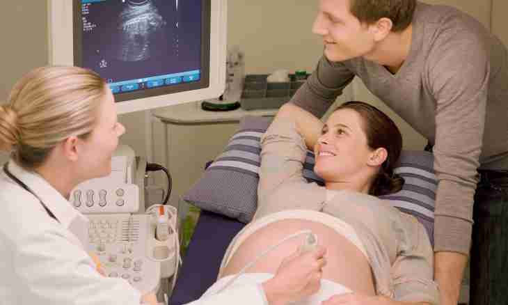
Ultrasonography or ekhografiya is considered today in almost only rather informative and rather safe way which allows to observe and estimate pregnancy development from beginning to end.
The first ultrasonography at pregnancy is carried out to 10-14 weeks if there are no indications to earlier research. Then ultrasonography is carried out to 5-6 weeks. (5-6 weeks) to ultrasonography the following factors can be indications to early: - approach of pregnancy at uterus myoma; - existence of a kontatseptiv in a uterus; - signs of possible termination of pregnancy (bloody allocations, pains in a stomach bottom); - suspicion on the "stood" or extra-uterine pregnancy. During the first ultrasonography the obstetrician-gynecologist can use two methods of carrying out inspection. Transvaginal research. It is conducted through a vagina. Advantage is that such ultrasonography gives fuller information on a condition of a fruit and a uterus since sensors approach closer to the studied objects and also have the big frequency of radiation. This method is used most often in the first trimester and the more so at ultrasonography on the earliest terms (5-6 weeks). Transabdominal research. It is carried out through an anterior abdominal wall. To such research it is necessary to be ready, namely, to have a full bladder. Ultrasonography of the first trimester pursues itself the following aims: - determination of arrangement of fetal egg (uterine or extra-uterine pregnancy); - diagnosing of polycarpous pregnancy; - measurement of growth and the structure of an embryo and assessment of the obtained data; - diagnostics of possible complications (the menacing abortion); - researches regarding definition or an exception of malformations and diseases (a tumor, malignant and good-quality, cysts, pathology of the structure of a uterus and so forth).