Heart is powerful muscular body which forces blood via the cameras and valves in the distributive system also known as the blood circulatory system. Heart near the center of a chest cavity is located.
General information
The science cardiology is engaged in studying heart. The average mass of heart is 250-300 grams. Heart has the cone-shaped form. Is its part, generally strong elastic fabric – the cardiac muscle which is rhythmically reduced throughout all life and overtaking through arteries and capillaries to body tissues blood. Average frequency of reductions of heart – about 70 times a minute.
Departments of heart
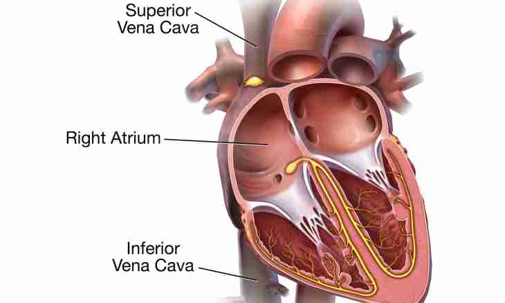
Heart of the person by partitions is divided into four cameras which at different times are filled with blood. The lower thick-walled chambers of heart are called ventricles. They act as the pump and after receiving blood from the top cameras by reduction send it to arteries. Process of reduction of ventricles is and there is heartbeat. The top cameras are called auricles which thanks to elastic walls easily stretch and contain in themselves the blood arriving between reductions from veins.
The left and right departments of heart are separated from each other, each of them consists of an auricle and a ventricle. The blood poor in oxygen flowing from body tissues at first comes to the right department, and later goes to lungs. The oxygenated blood from lungs comes to the left department, on the contrary, and it is redirected to all tissues of a body. For the reason that the left ventricle performs the most hard work which consists in forcing of blood on a big circle of blood circulation, it differs from other cameras of heart in the massiveness and bigger wall thickness – nearly 1.5 cm. In each half of heart of an auricle and ventricles are connected among themselves by the opening closed by the valve. Valves open only towards ventricles. This process is helped by the tendinous threads which are attached one end to shutters of valves, and opposite to the sosochkovy muscles which are placed on walls of ventricles. Such muscles represent outgrowths of a wall of ventricles and are reduced along with them, bringing tendinous threads into a tension and not passing blood back in an auricle. Tendinous threads prevent an eversion of valves towards auricles at reduction of ventricles. In places where the aorta leaves the left ventricle, and a pulmonary artery - from the right ventricle, semi-lunar valves in the form of pockets are placed. Blood passes through them in an aorta and a pulmonary artery, however the movement back in ventricles is impossible because semi-lunar valves when filling with blood are straightened and closed.